Description
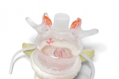
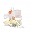
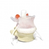
The Best Way To “Show” Decompression. Life-Like Lumbar Disc Model
New features include:
- circumferential (diffuse) disc bulge
- superimposed disc protrusion
- limacon shaped annulus
- peripherally exposed calcified endplate
- elastomeric white articular cartilage
- subchondrial bone exposed with hyaline fibrillation
- bone coloured L5
- white superior endplate matching the colour of the articular cartilage
- New cauda equina
The features that remain (from the previous model):
- flexible and totally dynamic herniating (or prolapse) nucleus pulposus. This is achieved through a realistic 2-part intervertebral disc with 6 degrees of freedom. Nuclear migration upon manual compression through a torn annulus fibrosus explaining pain generators under load.
- right posterior-lateral radial and circumferential(concentric) fissure
- transparent L4
- randomly scattered and embedded black nuclear structures to easily show nuclear shifting dynamics through the L4 view lens
- L5 superior endplate pores (black)
- L5 superior endplate lesion (red)
- vasculature in L4 vertebral body (red)
- facet subchondrial vascularization (red)
- facet tropism (L5 inferior)
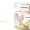
- Detailed cauda equina includes: sensory and motor divisions, dorsal root ganglion, recurrent meningeal, gray rami communicans, posterior primary division, dura mater, arachnoid sheath, rootlets, properly placed nerve root to accurately demonstrate the most commonly affected nerve with a post-lateral herniated disc.
Be the first to review “Dynamic Disc Model” Cancel reply
Related products

 Adjustable Axilla Posts (Set) * KDT TABLE OWNERS ONLY




Reviews
There are no reviews yet.